 my liking.
Anyway, enough whining! To the right here we have a photo at 400x mag (oil) of a retina showing GFAP+ astrocytes in the (green) in the nerve fibre layer wrapped around blood vessel (red/yellow). Although I'm not particularly enamoured of this photo because the yellow indicates that something may have gone wrong with the colours, I do like how the shape of the cell lining the blood vessel (pericyte) comes up clearly - the little round thing is where the nucleus would be if I threw a nuclear stain on. But this is like taking a whole salami and just cutting it out to see where the fat is.
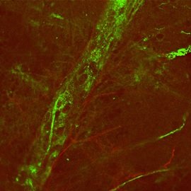 This (very bad) photo on the left is a photo of what the whole salami would look like if we could make it kinda transparent! This is a whole mounted retina with the blood vessel (green) surrounded by individual astrocytes (red). Ugh, this photo is sooo bad, I'm kinda disgusted at my inability to have made this experiment and the photography better *sigh*. Next time, I better make sure I mount the retina the right way up - I put it on the slide upside-down so when I took the photo, I had to focus through all the stupid layers of the retina! Add to that the fact that the retina I was using kind fell apart during the exp...I suppose I should be happy that I managed to get something like this at all.
No comments:
Post a Comment