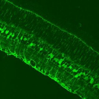
Anyway, I finally got the photography machine to spit some data out at me! It took three hours sitting in a hot dark room, but I FINALLY got some lovely photos! Here are some!
 This photo shows a vertical section of cryo-frozen retina stained for S100beta, a protein involved in calcium homeostasis and is produced by glial cells in the CNS. This slide is showing the cell bodies and processes of astrocyte cells at the bottom of the pic, Muller cell bodies and processes in the centre, and the Inner Limiting Membrane formed by MC endfeet at the top.
(1:50000, 400x mag)
 Hmm....lost a bit of the fine detail when I compressed this. This is a flatmounted whole retina labelled for EAAT-4, a glutamate transporter only expressed on astrocytes in the retina. So what the bright red things are are astrocyte processes. This doesnt look as nice as it does as a .tif file. (1:100, 630x mag)
So there's the proof that I don't really spend all day at uni on msn :P
No comments:
Post a Comment